
- 06 Aug 2013 00:52
#14284075
The thread title is literal.
I have rather good vision, but it seems that it is hardly normal and there are other physiological and cognitive factors associated as well. The data is very limited, as to the number of people who share these traits.
So, I am curious about how many here on the forum, might share one, or more of said traits.
Note: I will do some basic explanations/presentation then plan on providing more in depth data/related information at the end.
Here we go.
When most people close their eyes they see this:
Just a dark empty space and sometimes there might be a few specs of light, but predominately there is nothing but darkness.
This is called Eigengrau aka 'dark light' and is explained further here.
I see this,

but there are many different colors and more intricacies in the shapes. Sort of like this:

Something in-between is closer to what I see and the last one is real close. (With concentration I can also alter the shapes and colors, mostly blue)
Anyway,
This is called closed-eye visualizations (CEV), yet there are differences, as I have no physical, or psychological ailments.
This phenomenon is also known as phosphene, it is characterized by the experience of seeing light, without light actually entering the eye. Phosphenes are flashes of light, often associated with optic neuritis, induced by movement or sound, but I do not suffer from this.
This has also been called Prisoner's cinema. I won't go into it, as it's repetitive, but I like corroboration.
What I see is much more colorful and as I describe above. The article gives an example of this would look like, I show it below, but close and rub your eyes and you will get a good idea. (I do not think the below images is even close, at least for me)
This has also been attributed to other ailments and or drugs and I do not use and have no health problems.
The above description mentions another part of what I see, but is also uncommon.

I see this: However, for me, the the snow looks more like pixels and the image is clear and crisp, it just also has a active portion to the visual field.

This is called, Visual snow.
Another name is Hallucinogen persisting perception disorder, and though I see all the visual aspects described...
I have not used these type of drugs, or any prescriptions that would cause the disorder, nor do I have any of the physical/mental/neurological ailments associated with this disorder.
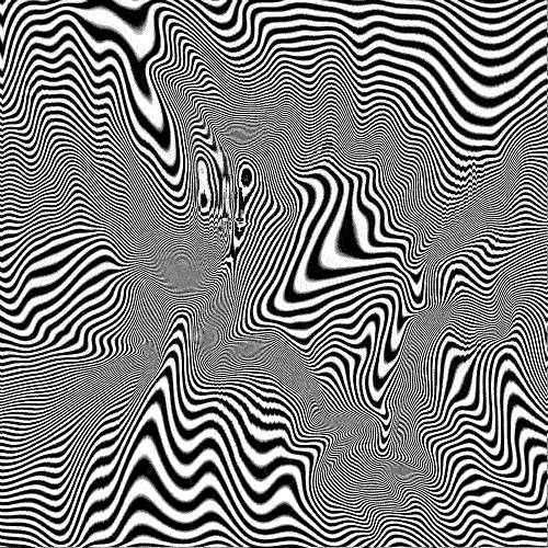
I also see what some call trailers and that looks like this:

From what I have read what I am seeing is not the same type of trailers that one might see while using drugs. Nor is what I am seeing just in my mind, that is to say that I perceive reality differently.
I say this because, the trailers allow me to track the trajectory of movement, unlike the hand above. I see this,

This also allows me to judge where the hand will end its movement. Now this could be just me learning to put what I am seeing to good use, but it goes past that.
Let me explain, looking at the image below and you can see the heat waves coursing out and away from the jet. Once the jet goes further away most people no longer see this air being moved.
I do. I also see those little particles I described above, moving away further and further through the air. The best description I can think of is, that I am seeing air current.

I have shown others by, seeing the air flow from a door, or window into the home and then describe that pattern, how the air moves, bounces off this, etc. Then I can show others that this is happening by just blowing smoke into the air.
As for visual there is only one other thing and it does fall under cognitive differences.
If you can see these, most see this:

I see this:

This would mean that my receptors do not flip this images. I have yet to find good information regarding this.
I have noticed that I see both sides of an optical illusion at the same time, so I see both the duck and the bunny instantly and this is the same for all similar images.

Anyway this ends the 'Do you see, what I see portion'. Past this point I share the research I have done on the subject.
I touch on dynamic relationships between vision, perception, memory, learning, sleep patterns, pain threshold and advanced physiology/psychology, in regards to the above information that I have shared.
I do come to a conclusion, so the post is not just ramblings.
_______________________________________________________________________________________________________________
From what I have found, I believe that all of the above can be attributed to the neurological response to light. This then starts with the pupillary response.

I will come back to this subject later.
I have a visual memory and very good recall, so I can't help thinking that this is associated with classical conditioning in regards to how I sort and store visual input and that the increases in said recall are associated with said classical conditioning.
I will come back to visual memory later on.
Visual information goes to the retina, then the thalamus, and then to the primary visual cortex. I could not see how the retina or the thalamus could be responsible for such alterations to the normal human condition so I moved on to looking at the visual cortex.
I found very early on that I could control my pupillary response voluntarily and often wonder if that control interrupted the bodies natural ordering of the visual association cortex.
Information received by the visual cortex is sent 'maps' of the visual field within the visual association cortex.
This then is our perception. I then wonder if that alter perception is than reflected back to withing the visual filed.
Damage to the brain can effect the visual association cortex and this can alter ones perception by messing with ones Form perception. Normally this type of injury is also associated with interfering with the ability to see motions and colors.
(I have taken some nasty blows but have have never been any worse for the ware. Besides the fact that I have always been this way. )
So from this I assume that the visual differences that I am seeing are associated with the enhancement of the visual association cortex and specifically the processing of input in regards to Form perception. This would account for the enhanced colors, motion tracking and the ability to perceive minute changes in the atmosphere.
The last assumedly because the atmospheric particles are being illuminated more.
This part intrigues me, as it would make sense that this portion is why I see the magic eye images inverted.
Back to visual memory. Besides for the definition provided below, I wanted to add that for me, my recall includes getting a visual of the item, face pattern, etc. To the point that I see the page and can read the information that I am seeking from the visual memory of the page.

The reason that I am looking at this, is because of the association with the visual cortex and the fact that it seems that I am hard wired in a different way that is dominated by visual responses.
So again this brings me back to the pupillary response.
In this case I look at the pupillary responses interaction with the circadian rhythm.
(I have a rather odd sleep schedule and need very little sleep, 4 hours a day is often enough and I can stay up for extremely long periods with no cognitive losses)
I was able to discern that my sleep pattern placed me within a Non-24-hour sleep-wake disorder. However, I still do not suffer from any of the symptoms that are normally associated with the disorder.
I found this interesting because the majority of those who have this disorder are blind.
Here I noticed that the Optic chiasm is mentioned and that it controls the aspect where ones sees the left/right cross over. This aspect being affected would explain why I see the duck and the bunny at the same time.
So I traveled down that road.
What I got out of researching this area is that the reticular activating system and the ganglion cells control this portion separate from the thalamus and that the relay is a back and forth informational system through the Superior colliculus.
This would keep in holding with the idea that my Form perception is being bounce back to my visual cortex in an amplified manner. The question was why.
The answer comes in the area of optic radiation through the Superior colliculus and the ganglion cells of the retina.
So it is becoming apparent that, for whatever reason, my body has no, or less distinction between these superficial layers.
The problem I ran into is that Primates no longer have this functionality of the brain.
Accordingly I found the problem not to be in the parabigeminal nucleus but instead it must be in the Pretectal area. I take one look and think bingo, but of course I want more than the basic anatomical connection, as my condition is not normal and apparently not supposed to be happening.
So here this area connects all the above aspects, the subcortical visual system, the projections to the retina, pupillary light reflex, acute changes in ambient light and changes to my circadian rhythm.
On a side note this explains my extremely high tolerance of pain. Except that is the opposite described, go figure.
So I narrow all of this down to the Anterior Pretectal Nucleus...
and further to the dorsal horn. Shown below.


This takes me right to Photosensitive ganglion cells.
So at last it seems I found why I see the way I do.
You can read the whole article here named: Strange vision.
I have rather good vision, but it seems that it is hardly normal and there are other physiological and cognitive factors associated as well. The data is very limited, as to the number of people who share these traits.
So, I am curious about how many here on the forum, might share one, or more of said traits.
Note: I will do some basic explanations/presentation then plan on providing more in depth data/related information at the end.
Here we go.
When most people close their eyes they see this:
Just a dark empty space and sometimes there might be a few specs of light, but predominately there is nothing but darkness.
This is called Eigengrau aka 'dark light' and is explained further here.
I see this,
but there are many different colors and more intricacies in the shapes. Sort of like this:
Something in-between is closer to what I see and the last one is real close. (With concentration I can also alter the shapes and colors, mostly blue)
Anyway,
This is called closed-eye visualizations (CEV), yet there are differences, as I have no physical, or psychological ailments.
This phenomenon is also known as phosphene, it is characterized by the experience of seeing light, without light actually entering the eye. Phosphenes are flashes of light, often associated with optic neuritis, induced by movement or sound, but I do not suffer from this.
This has also been called Prisoner's cinema. I won't go into it, as it's repetitive, but I like corroboration.
The most common phosphenes are pressure phosphenes, caused by rubbing the closed eyes. The pressure mechanically stimulates the cells of the retina. Experiences include a darkening of the visual field that moves against the rubbing, a diffuse colored patch that also moves against the rubbing, a scintillating and ever-changing and deforming light grid with occasional dark spots (like a crumpling fly-spotted flyscreen), and a sparse field of intense blue points of light. Pressure phosphenes can persist briefly after the rubbing stops and the eyes are opened, allowing the phosphenes to be seen on the visual scene.
What I see is much more colorful and as I describe above. The article gives an example of this would look like, I show it below, but close and rub your eyes and you will get a good idea. (I do not think the below images is even close, at least for me)
This has also been attributed to other ailments and or drugs and I do not use and have no health problems.
The above description mentions another part of what I see, but is also uncommon.
Pressure phosphenes can persist briefly after the rubbing stops and the eyes are opened, allowing the phosphenes to be seen on the visual scene.That being the last part, when I am looking around I do not see this:
I see this: However, for me, the the snow looks more like pixels and the image is clear and crisp, it just also has a active portion to the visual field.
This is called, Visual snow.
Visual snow is a transitory or persisting visual symptom where people see snow or television-like static in parts or the whole of their visual fields, especially against dark backgrounds. It is much like camera noise in low light conditions.
The severity or density of the "snow" differs from one person to the next; in some circumstances, it can inhibit a person's daily life, making it difficult to read, see in detail and focus correctly. The "snow" is more generalized than the "blue-sky sprites" or "worms" seen in the blue field entoptic phenomenon.
No etiology for visual snow has been identified, and anecdotal reports point to a multitude of associated conditions, possibly rendering it a non-specific symptom. Insofar as sufferers of visual snow have undergone ophthalmic, neurological and psychiatric examinations, no systematic problems besides the visual snow have been identified. Pending recognition of the condition, little medical research is taking place to possibly identify more subtle deviations.
Another name is Hallucinogen persisting perception disorder, and though I see all the visual aspects described...
There are a number of perceptual changes that can accompany HPPD. Typical symptoms of the disorder include: halos or auras surrounding objects, trails following objects in motion, difficulty distinguishing between colors, apparent shifts in the hue of a given item, the illusion of movement in a static setting, air assuming a grainy or textured quality (visual snow or static, by popular description, not to be confused with normal "blue field entoptic phenomenon"), distortions in the dimensions of a perceived object, and a heightened awareness of floaters. The visual alterations experienced by those with HPPD are not homogeneous and there appear to be individual differences in both the number and intensity of symptoms.
I have not used these type of drugs, or any prescriptions that would cause the disorder, nor do I have any of the physical/mental/neurological ailments associated with this disorder.
I also see what some call trailers and that looks like this:
From what I have read what I am seeing is not the same type of trailers that one might see while using drugs. Nor is what I am seeing just in my mind, that is to say that I perceive reality differently.
I say this because, the trailers allow me to track the trajectory of movement, unlike the hand above. I see this,
This also allows me to judge where the hand will end its movement. Now this could be just me learning to put what I am seeing to good use, but it goes past that.
Let me explain, looking at the image below and you can see the heat waves coursing out and away from the jet. Once the jet goes further away most people no longer see this air being moved.
I do. I also see those little particles I described above, moving away further and further through the air. The best description I can think of is, that I am seeing air current.
I have shown others by, seeing the air flow from a door, or window into the home and then describe that pattern, how the air moves, bounces off this, etc. Then I can show others that this is happening by just blowing smoke into the air.
As for visual there is only one other thing and it does fall under cognitive differences.
If you can see these, most see this:
I see this:
This would mean that my receptors do not flip this images. I have yet to find good information regarding this.
I have noticed that I see both sides of an optical illusion at the same time, so I see both the duck and the bunny instantly and this is the same for all similar images.
Anyway this ends the 'Do you see, what I see portion'. Past this point I share the research I have done on the subject.
I touch on dynamic relationships between vision, perception, memory, learning, sleep patterns, pain threshold and advanced physiology/psychology, in regards to the above information that I have shared.
I do come to a conclusion, so the post is not just ramblings.
_______________________________________________________________________________________________________________
From what I have found, I believe that all of the above can be attributed to the neurological response to light. This then starts with the pupillary response.
Pupillary response is a physiological response that varies the size of the pupil,via the trigeminal cranial nerve, either resulting in constriction (miosis)[1] or dilation (mydriasis, expansion), via relaxation or contraction of the iris dilator muscle. The response can have a variety of causes, from an involuntary reflex reaction to exposure or inexposure to light — in low light conditions a dilated pupil lets more light into the eye — or it may indicate interest in the subject of attention or indicate sexual stimulation.[2] The pupils contract immediately before someone falls asleep.[3] A pupillary response can be intentionally conditioned as a Pavlovian response to some stimuli
I will come back to this subject later.
I have a visual memory and very good recall, so I can't help thinking that this is associated with classical conditioning in regards to how I sort and store visual input and that the increases in said recall are associated with said classical conditioning.
I will come back to visual memory later on.
"In any animal, regardless of its prior history, painful stimulation of the foot causes the leg to be withdrawn by bending at all its joints. This flexor reflex is an example of an unconditioned reflex, an innate response based on fixed connections in the chain of neurons from the receptor (sensor) to the effector. Of still more interest in everyday life are the acquired or conditioned reflexes, in which the functional connections between the excited sensors and the patterns of activity in effector organs become established by learning process."
Visual information goes to the retina, then the thalamus, and then to the primary visual cortex. I could not see how the retina or the thalamus could be responsible for such alterations to the normal human condition so I moved on to looking at the visual cortex.
I found very early on that I could control my pupillary response voluntarily and often wonder if that control interrupted the bodies natural ordering of the visual association cortex.
Neurons that are interposed between the afferent and efferent limbs of this circuit and include the visual association cortex, which determines the image is "out-of-focus, ends corrective signals via the internal capsule and crus cerebri to the supraoculomotor nuclei. It also includes the supraoculomotor nuclei (located immediately superior to the oculomotor nuclei) that generates motor control signals that initiate the accommodation response and sends these control signals bilaterally to the oculomotor complex.
Information received by the visual cortex is sent 'maps' of the visual field within the visual association cortex.
This then is our perception. I then wonder if that alter perception is than reflected back to withing the visual filed.
Damage to the brain can effect the visual association cortex and this can alter ones perception by messing with ones Form perception. Normally this type of injury is also associated with interfering with the ability to see motions and colors.
(I have taken some nasty blows but have have never been any worse for the ware. Besides the fact that I have always been this way. )
Form perception is the recognition of objects in the environment. We originally perceive an object on our retina as a 2D image.[1] The Retinal image can vary for the same object in terms of the context with which it is viewed, the apparent size of the object, the angle from which it is viewed, how illuminated it is, as well as where it resides in our field of vision.
So from this I assume that the visual differences that I am seeing are associated with the enhancement of the visual association cortex and specifically the processing of input in regards to Form perception. This would account for the enhanced colors, motion tracking and the ability to perceive minute changes in the atmosphere.
The last assumedly because the atmospheric particles are being illuminated more.
Through visual processing in the temporal, parietal and occipital lobe these retinal images are converted into a 3D image
This part intrigues me, as it would make sense that this portion is why I see the magic eye images inverted.
Back to visual memory. Besides for the definition provided below, I wanted to add that for me, my recall includes getting a visual of the item, face pattern, etc. To the point that I see the page and can read the information that I am seeking from the visual memory of the page.
Visual memory describes the relationship between perceptual processing and the encoding, storage and retrieval of the resulting neural representations. Visual memory occurs over a broad time range spanning from eye movements to years in order to visually navigate to a previously visited location.[1] Visual memory is a form of memory which preserves some characteristics of our senses pertaining to visual experience. We are able to place in memory visual information which resembles objects, places, animals or people in a mental image. The experience of visual memory is also referred to as the mind's eye through which we can retrieve from our memory a mental image of original objects, places, animals or people.[1] Visual memory is one of several cognitive systems, which are all interconnected parts that combine to form the human memory.[2]
The reason that I am looking at this, is because of the association with the visual cortex and the fact that it seems that I am hard wired in a different way that is dominated by visual responses.
The visual cortex also receives information from subcortical regions, such as the lateral geniculate body, located in the thalamus. However, ample evidence indicates that object identity and location are preferentially processed in ventral (occipito-temporal) and dorsal (occipito-parietal) cortical visual streams, respectively.[3] Comparison of rCBF during performance of the two tasks again revealed differences between the ventral and dorsal pathways.
So again this brings me back to the pupillary response.
In this case I look at the pupillary responses interaction with the circadian rhythm.
(I have a rather odd sleep schedule and need very little sleep, 4 hours a day is often enough and I can stay up for extremely long periods with no cognitive losses)
A circadian rhythm /sɜrˈkeɪdiən/ is any biological process that displays an endogenous, entrainable oscillation of about 24 hours. These rhythms are driven by a circadian clock, and rhythms have been widely observed in plants, animals, fungi and cyanobacteria. The term circadian comes from the Latin circa, meaning "around" (or "approximately"), and diem or dies, meaning "day". The formal study of biological temporal rhythms, such as daily, tidal, weekly, seasonal, and annual rhythms, is called chronobiology. Although circadian rhythms are endogenous ("built-in", self-sustained), they are adjusted (entrained) to the local environment by external cues called zeitgebers, commonly the most important of which is daylight.
I was able to discern that my sleep pattern placed me within a Non-24-hour sleep-wake disorder. However, I still do not suffer from any of the symptoms that are normally associated with the disorder.
In people with this disorder, the body essentially insists that the day is longer than 24 hours and refuses to adjust to the external light/dark cycle. This makes it impossible to sleep at normal times and also causes daily shifts in other aspects of the circadian rhythms such as peak time of alertness, body temperature minimum and hormone secretion. Left untreated, non-24-hour sleep-wake syndrome causes a person's sleep-wake cycle to change every day, the degree determined by how much over 24 hours the cycle lasts. The cycle progresses around the clock, eventually returning to "normal" for one or two days before "going off" again.
I found this interesting because the majority of those who have this disorder are blind.
Sighted people with Non-24 do exist, but are much more rare and the etiology of their circadian disorder is less well understood.[2] As of 2005, there were fewer than 100 cases of sighted people with N24 reported in the scientific literature.[8] At least one case of a sighted person developing Non-24 was preceded by head injury;[9] another patient diagnosed with the disorder was later found to have a "large pituitary adenoma that involved the optic chiasma".[1] Thus the problem appears to be neurological. Specifically, it is thought to involve abnormal functioning of the suprachiasmatic nucleus of the hypothalamus.[10] Several other cases have been preceded by chronotherapy, a prescribed treatment for delayed sleep phase syndrome.[2][11]
There have been several studies of sighted people with the disorder. McArthur et al. reported treating a sighted patient who "appeared to be subsensitive to bright light".[12] In other words, the brain does not react normally to light (people with the disorder may or may not, however, be unusually subjectively sensitive to light; one study found that they were more sensitive than the control group.[13]) In 2002 Uchiyama et al. examined five sighted Non-24 patients who showed, during the study, a sleep-wake cycle averaging 25.12 hours.[14] That is appreciably longer than the 24.02-hour average shown by the control subjects in that study, which was near the average innate cycle for healthy adults, younger and older, of 24.18 hours.[15] The literature usually refers to a "one to two hour" delay per 24-hour day (i.e. a 25–26 hour cycle).
Here I noticed that the Optic chiasm is mentioned and that it controls the aspect where ones sees the left/right cross over. This aspect being affected would explain why I see the duck and the bunny at the same time.
So I traveled down that road.
Beyond the optic chiasm, with crossed and uncrossed fibers, the optic nerves become optic tracts. The signals are passed on to the lateral geniculate body, in turn giving them to the occipital cortex (the outer matter of the rear brain)
The lateral geniculate nucleus (LGN) is the primary relay center for visual information received from the retina of the eye. The LGN is found inside the thalamus of the brain. The LGN receives information directly from the ascending retinal ganglion cells via the optic tract and from the reticular activating system. Neurons of the LGN send their axons through the optic radiation, a direct pathway to the primary visual cortex. In addition, the LGN receives many strong feedback connections from the primary visual cortex.
What I got out of researching this area is that the reticular activating system and the ganglion cells control this portion separate from the thalamus and that the relay is a back and forth informational system through the Superior colliculus.
This would keep in holding with the idea that my Form perception is being bounce back to my visual cortex in an amplified manner. The question was why.
The answer comes in the area of optic radiation through the Superior colliculus and the ganglion cells of the retina.
The microstructure of the optic tectum / superior colliculus varies across species. As a general rule, there is always a clear distinction between superficial layers, which receive input primarily from the visual system and show primarily visual responses, and deeper layers, which receive many types of input and project to numerous motor-related brain areas. The distinction between these two zones is so clear and consistent that some anatomists have suggested that they should be considered separate brain structures.
So it is becoming apparent that, for whatever reason, my body has no, or less distinction between these superficial layers.
The superficial layers receive input mainly from the retina, vision-related areas of the cerebral cortex, and two tectal-related structures called the pretectum and parabigeminal nucleus. The retinal input encompasses the entire superficial zone, and is bilateral, although the contralateral portion is more extensive. The cortical input comes most heavily from the primary visual cortex (area 17), the secondary visual cortex (areas 18 and 19), and the frontal eye fields.
The parabigeminal nucleus plays a very important role in tectal function that will be described below.
In contrast to the vision-dominated inputs to the superficial layers, the intermediate and deep layers receive inputs from a very diverse set of sensory and motor structures. Most areas of the cerebral cortex project to these layers, although the input from "association" areas tends to be heavier than the input from primary sensory or motor areas.[citation needed] However, the cortical areas involved, and the strength of their relative projections differs across species.[12] Another important input comes from the substantia nigra, pars reticulata, a component of the basal ganglia. This projection uses the inhibitory neurotransmitter GABA, and is thought to exert a "gating" effect on the superior colliculus. The intermediate and deep layers also receive input from the spinal trigeminal nucleus, which conveys somatosensory information from the face, as well as the hypothalamus, zona incerta, thalamus, and inferior colliculus.
The problem I ran into is that Primates no longer have this functionality of the brain.
It is usually accepted that the primate superior colliculus is unique among mammals, in that it does not contain a complete map of the visual field seen by the contralateral eye. Instead, like the visual cortex and lateral geniculate nucleus, each colliculus represents only the contralateral half of the visual field, up to the midline, and excludes a representation of the ipsilateral half.[25] This functional characteristic is explained by the absence, in primates, of anatomical connections between the retinal ganglion cells in the temporal half of the retina and the contralateral superior colliculus. In other mammals, the retinal ganglion cells throughout the contralateral retina project to the contralateral colliculus.
Accordingly I found the problem not to be in the parabigeminal nucleus but instead it must be in the Pretectal area. I take one look and think bingo, but of course I want more than the basic anatomical connection, as my condition is not normal and apparently not supposed to be happening.
The pretectal area, or pretectum, is a midbrain structure composed of seven nuclei and comprises part of the subcortical visual system. Through reciprocal bilateral projections to the retina, it is primarily involved in mediating behavioral responses to acute changes in ambient light such as the pupillary light reflex, the optokinetic reflex and temporary changes to the circadian rhythm.[1][2][3][4][5] In addition to the pretectum's role in the visual system, the anterior pretectal nucleus has been found to mediate somatosensory and nociceptive information
So here this area connects all the above aspects, the subcortical visual system, the projections to the retina, pupillary light reflex, acute changes in ambient light and changes to my circadian rhythm.
The pretectum is a bilateral group of highly interconnected nuclei located near the junction of the midbrain and forebrain.[8] The pretectum is generally classified as a midbrain structure, although because of its proximity to the forebrain it is sometimes classified as part of the caudal diencephalon (forebrain).[9] Within vertebrates, the pretectum is located directly anterior to the superior colliculus and posterior to the thalamus. Vertically, it is situated above the periaqueductal grey and nucleus of the posterior commissure.[10]
Several nuclei have been identified within the pretectum, although their borders can be difficult to define and there has been debate over which regions should be included and their precise names.[1][10][11] The five primary nuclei are: the olivary pretectal nucleus (ON), the nucleus of the optic tract (NTO), and the anterior (NPA), medial (NPM), and posterior (NPP) pretectal nuclei. The NTO consists of relatively large cells and is located between the superior colliculi. The OPN is located medial to the NTO and has a tail that extends between the NTO and NPP, which is ventral to the ON.[10] Two additional nuclei have also been identified: the posterior limitans (PLi) and the commisural pretectal area (CPA).[12] While these two regions have not been examined to the same extent as the five primary nuclei, research has shown both the PLi and CPA to receive retinal input which suggests a role in processing visual information.[13]
Inputs
The pretectum receives significant binocular input from photosensitive ganglion cells in the retina. In primates these afferents are bilateral[14] while in rodents they project from the contralateral retina. The majority of these retino-pretectal projections go to the ON and NTO[14] while other pretectal nuclei receive minor retinal input in mammals including the posterior, medial, and anterior pretectal nuclei.[15][1][10][16]
The NTO receives input from several regions. From the thalamus the NTO receives inhibitory projections from GABA producing neurons in the ipsilateral lateral geniculate nucleus and bilateral intergeniculate leaflets. The ipsilateral superficial suprachiasmatic nucleus and the medial, dorsal, and lateral terminal nuclei in the midbrain project onto the NTO. Fibers also project to the NTO from the nucleus prepositus hypoglossi in the medulla, the contralateral NTO, and from various cortical regions.[1][17]
Outputs
Many pretectal nuclei share targets of efferent projections. All pretectal nuclei, except for the ON, project to nuclei in the thalamus, subthalamus, superior colliculus, reticular formation, pons, and inferior olive.[10] Both the ON and the CPA have efferent projections to the Edinger-Westphal nucleus. The NPP and NPA both project to the pulvinar, the lateral posterior nucleus of the thalamus, and several precerebellar nuclei.[1]
The NTO has efferent projections to the zona incerta of the subthalamus, several nuclei of the pons, medulla, intralaminar nuclei, midbrain, and dorsal and ventral thalamic nuclei. Its bilateral inhibitory projections to the accessory optic system include connections to the lateral and medial terminal nuclei. Projections to the subthalamus are target towards the lateral geniculate nucleus and pulvinar. The NTO projects bilaterally to the superior colliculus, although the ipsilateral connections appear to be more dominant. In addition to these projections, the NTO projects to the vestibular and vestibulocerebellar relay nuclei.[1]
On a side note this explains my extremely high tolerance of pain. Except that is the opposite described, go figure.
The NPA participates in the active diminishing of the perception of pain stimuli (antinociception).[7] Although the mechanism by which the NPA alters an organism's response to painful stimuli is not fully known, research has shown that activity in the ventral NPA triggers cholinergic and serotonergic neurons. These neurons activate descending pathways that synapse in the spinal cord and inhibit nociceptive cells in the dorsal horn.[22][23] In addition to its direct antionociceptive mechanism, the NPA projects onto brain regions that, through connections to the somatosensory cortex, regulate the perception of painful stimuli. Two of these regions that the NPA is known to project to are the zona incerta and posterior thalamic nucleus. Regions of the NPA may be specialized to respond to different types of pain. Research has found that the dorsal NPA best diminished the perception of brief pain whereas the ventral NPA reduced the perception of chronic pain.[24] Because of its role in the reduction of chronic pain, abnormal activity of the NPA is thought to be implicated in central pain syndrome.
So I narrow all of this down to the Anterior Pretectal Nucleus...
Multireceptive neurones deep in the dorsal horn are inhibited by APtN stimulation. In contrast, superficial projection neurones that respond to intense cutaneous stimuli are excited by APtN stimulation. The APtN receives an excitatory input from low-threshold afferents via the dorsal column pathway and a high-threshold excitatory drive from superficial cells projecting through the dorsolateral funiculus. The excitatory input from the dorsal columns may well participate in the long-term inhibition of spinal projection neurones evoked by dorsal column stimulation. These ascending excitatory pathways may also be important to the long-term activation of descending inhibition from the APtN.
and further to the dorsal horn. Shown below.
This takes me right to Photosensitive ganglion cells.
Photosensitive ganglion cells, also called photosensitive Retinal Ganglion Cells (pRGC), intrinsically photosensitive Retinal Ganglion Cells (ipRGC) or melanopsin-containing ganglion cells, are a type of neuron (nerve cell) in the retina of the mammalian eye. They were discovered in 1923,[1] forgotten, rediscovered in the early 1990s[2] and are, unlike other retinal ganglion cells, intrinsically photosensitive. This means that they are a third class of retinal photoreceptors, excited by light even when all influences from classical photoreceptors (rods and cones) are blocked (either by applying pharmacological agents or by dissociating the ganglion cell from the retina). Photosensitive ganglion cells contain the photopigment melanopsin. The giant retinal ganglion cells of the primate retina are examples of photosensitive ganglion cells.
Compared to the rods and cones, the ipRGC respond more sluggishly and signal the presence of light over the long term.[3] They represent a small subset (~1-3%) of the retinal ganglion cells. Their functional roles are non-image-forming and fundamentally different from those of pattern vision; they provide a stable representation of ambient light intensity. They have at least three primary functions.
They play a major role in synchronizing circadian rhythms to the 24-hour light/dark cycle, providing primarily length-of-day and length-of night information. They send light information via the retinohypothalamic tract directly to the circadian pacemaker of the brain, the suprachiasmatic nucleus of the hypothalamus. The physiological properties of these ganglion cells match known properties of the daily light entrainment (synchronization) mechanism regulating circadian rhythms.
Photosensitive ganglion cells innervate other brain targets, such as the center of pupillary control, the olivary pretectal nucleus of the midbrain. They contribute to the regulation of pupil size and other behavioral responses to ambient lighting conditions.
They contribute to photic regulation of, and acute photic suppression of, release of the hormone melatonin from the pineal gland.
Photosensitive ganglion cells are also responsible for the persistence of circadian and pupillary light responses in mammals with degenerated rod and cone photoreceptors, such as humans suffering from retinitis pigmentosa.
Recently photoreceptive ganglion cells have been isolated in humans where, in addition to the above functions shown in other mammals, they have been shown to mediate a degree of light recognition in rodless, coneless subjects suffering with disorders of rod and cone photoreceptors.[4] Work by Farhan H. Zaidi and colleagues showed that photoreceptive ganglion cells may have a visual function and can be isolated in humans.
The photopigment of photoreceptive ganglion cells, melanopsin, is excited by light mainly in the blue portion of the visible spectrum (absorption peaks at ~480 nanometers[5]). The phototransduction mechanism in these cells is not fully understood, but seems likely to resemble that in invertebrate rhabdomeric photoreceptors. Photosensitive ganglion cells respond to light by depolarizing and increasing the rate at which they fire nerve impulses. In addition to responding directly to light, these cells may receive excitatory and inhibitory influences from rods and cones by way of synaptic connections in the retina.
Possible role in conscious sight wrote:The use of rodless, coneless humans allowed another possible role for the receptor to be studied. In 2007, a new role was found for the photoreceptive ganglion cell. Zaidi and colleagues showed that in humans the retinal ganglion cell photoreceptor contributes to conscious sight as well as to non-image-forming functions like circadian rhythms, behaviour and pupillary reactions.[4] Since these cells respond mostly to blue light, it has been suggested that they have a role in mesopic vision[citation needed] and that the old theory of a purely duplex retina with rod (dark) and cone (light) light vision was simplistic. Zaidi and colleagues' work with rodless, coneless human subjects hence has also opened the door into image-forming (visual) roles for the ganglion cell photoreceptor.
The discovery that there are parallel pathways for vision was made - one classic rod and cone-based arising from the outer retina, the other a rudimentary visual brightness detector arising from the inner retina and which seems to be activated by light before the other.[4] Classic photoreceptors also feed into the novel photoreceptor system, and colour constancy may be an important role as suggested by Foster.
It has been suggested by the authors of the rodless, coneless human model that the receptor could be instrumental in understanding many diseases including major causes of blindness worldwide such as glaucoma, a disease which affects ganglion cells.
So at last it seems I found why I see the way I do.
You can read the whole article here named: Strange vision.
Strange vision wrote:A novel photoreceptor of the mammalian retina has
recently been discovered and characterized. The novel
cells differ radically from the classical rod and cone
photoreceptors. They use a unique photopigment,
most probably melanopsin. They have lower sensitivity
and spatiotemporal resolution than rods or cones and
they seem specialized to encode ambient light intensity.
Most surprisingly, they are ganglion cells and,
thus, communicate directly with the brain. These intrinsically
photosensitive retinal ganglion cells (ipRGCs)
help to synchronize circadian rhythms with the solar
day. They also contribute to the pupillary light reflex
and other behavioral and physiological responses to
environmental illumination.
Synchronization of circadian rhythms wrote:The roots of this discovery lie in the field of circadian
physiology. Circadian rhythms are biological cycles that
have period of about a day. Body temperature, hormonal
levels, sleep, cognitive performance and countless other
physiological variables exhibit such daily oscillations. In
mammals, a pacemaker in the hypothalamus called the
suprachiasmatic nucleus (SCN) drives these rhythms


 - By wat0n
 - By Negotiator
 - By litwin